The main arches are the antero-posterior arches, which may, for descriptive purposes, be regarded as divisible into two types�a medial and a lateral.
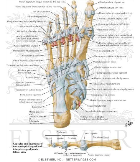
The medial arch is made up by the calcaneus, the talus, the navicular, the three cuneiforms, and the first, second, and third metatarsals.
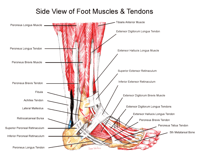
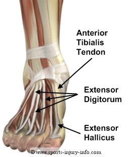
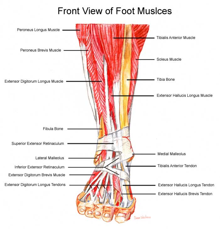
The arch is further supported by the plantar aponeurosis, by the small muscles in the sole of the foot, by the tendons of the Tibialis anterior and posterior and Peron�us longus, and by the ligaments of all the articulations involved.
At the posterior part of the metatarsus and the anterior part of the tarsus the arches are complete, but in the middle of the tarsus they present more the characters of half-domes the concavities of which are directed downward and medialward, so that when the medial borders of the feet are placed in apposition a complete tarsal dome is formed. The transverse arches are strengthened by the interosseous, plantar, and dorsal ligaments, by the short muscles of the first and fifth toes (especially the transverse head of the Adductor hallucis), and by the Peron�us longus, whose tendon stretches across between the piers of the arches.

Foot Trainer - Foot and Leg

tendons foot

the feet from moving in

foot: right foot, major

Muscles and Tendons

Ankle Tendons

The peroneal tendons (peroneus

Foot Tendonitis Diagram

Back of foot

Ligaments and Tendons of Foot:

three tendons that run the

tendons of the foot - the

Tendons are strong, fibrous

The foot is susceptible to

tendon in the foot.

Foot Tendonitis
The medial arch is made up by the calcaneus, the talus, the navicular, the three cuneiforms, and the first, second, and third metatarsals.
The arch is further supported by the plantar aponeurosis, by the small muscles in the sole of the foot, by the tendons of the Tibialis anterior and posterior and Peron�us longus, and by the ligaments of all the articulations involved.
At the posterior part of the metatarsus and the anterior part of the tarsus the arches are complete, but in the middle of the tarsus they present more the characters of half-domes the concavities of which are directed downward and medialward, so that when the medial borders of the feet are placed in apposition a complete tarsal dome is formed. The transverse arches are strengthened by the interosseous, plantar, and dorsal ligaments, by the short muscles of the first and fifth toes (especially the transverse head of the Adductor hallucis), and by the Peron�us longus, whose tendon stretches across between the piers of the arches.

Foot Trainer - Foot and Leg

tendons foot

the feet from moving in

foot: right foot, major

Muscles and Tendons

Ankle Tendons

The peroneal tendons (peroneus

Foot Tendonitis Diagram

Back of foot

Ligaments and Tendons of Foot:

three tendons that run the

tendons of the foot - the

Tendons are strong, fibrous

The foot is susceptible to

tendon in the foot.

Foot Tendonitis
No comments:
Post a Comment